What is Ultrasound?
What is medical ultrasound?
Cross-sectional ultrasound image of a fetus Source: Phillips Health Care – iu22xMATRIX system
Medical ultrasound is categorized into two primary types: diagnostic and therapeutic.
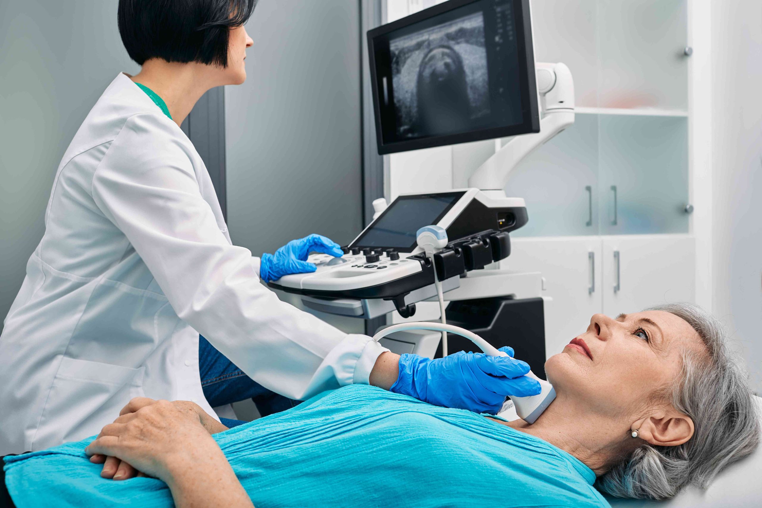
Diagnostic ultrasound is a non-invasive imaging technique that allows visualization of internal body structures. It employs ultrasound probes, known as transducers, which emit sound waves at frequencies exceeding the human hearing range (above 20 kHz). Most transducers currently in use operate at significantly higher frequencies, typically in the megahertz (MHz) range. While most diagnostic ultrasound probes are applied externally on the skin, they can also be inserted internally through the gastrointestinal tract, vagina, or blood vessels to enhance image quality. Additionally, ultrasound may be utilized during surgical procedures by introducing a sterile probe into the surgical site.
Diagnostic ultrasound can be further classified into anatomical and functional ultrasound. Anatomical ultrasound generates images of internal organs and structures, whereas functional ultrasound integrates data regarding tissue or blood movement, velocity, and physical properties such as softness or hardness with anatomical images to create “information maps.” These maps assist healthcare professionals in identifying functional changes or differences within a structure or organ.
Therapeutic ultrasound, on the other hand, also utilizes sound waves beyond the human hearing range but does not create images. Its primary aim is to interact with bodily tissues to modify or eliminate them. Potential modifications include moving or displacing tissue, heating it, dissolving blood clots, or delivering medications to targeted areas. The ablative capabilities of therapeutic ultrasound are facilitated by high-intensity beams that can eradicate diseased or abnormal tissues, such as tumors. A significant advantage of ultrasound therapy is that it is generally non-invasive, requiring no incisions or cuts, thus leaving no wounds or scars.
How Ultrsound Works?
Ultrasound waves are generated by a transducer, which functions to both emit these waves and detect the echoes that are reflected back. Typically, the active components of ultrasound transducers consist of specialized ceramic crystal materials known as piezoelectrics. These materials can generate sound waves when subjected to an electric field and can also reverse the process, creating an electric field in response to incoming sound waves. In the context of an ultrasound scanner, the transducer emits a beam of sound waves into the body. These waves are reflected back to the transducer by the interfaces between different tissues encountered along the beam’s path, such as the junctions between fluid and soft tissue or between tissue and bone. Upon receiving these echoes, the transducer converts them into electrical signals, which are transmitted to the ultrasound scanner. By measuring the speed of sound and the time taken for each echo to return, the scanner determines the distance from the transducer to the tissue boundary. This information is then utilized to create two-dimensional images of various tissues and organs.
During an ultrasound examination, the technician applies a gel to the skin. This gel prevents the formation of air pockets between the transducer and the skin, which could obstruct the passage of ultrasound waves into the body.
