Medical X-Ray Imaging
Advancements in medical imaging have significantly enhanced the diagnosis and treatment of various health conditions in both children and adults.
Medical imaging encompasses a range of procedures, known as modalities, each employing distinct technologies and techniques. Procedures such as computed tomography (CT), fluoroscopy, and radiography (commonly referred to as conventional X-ray, including mammography) utilize ionizing radiation to create images of the body. Ionizing radiation possesses sufficient energy to potentially harm DNA, which may increase an individual’s lifetime risk of developing cancer.
The fundamental principle behind CT, radiography, and fluoroscopy is similar: an X-ray beam is directed through the body, where some X-rays are absorbed or scattered by internal structures, while the remaining X-ray pattern is captured by a detector (such as film or a computer screen) for recording or further analysis by a computer. These imaging techniques serve different purposes:
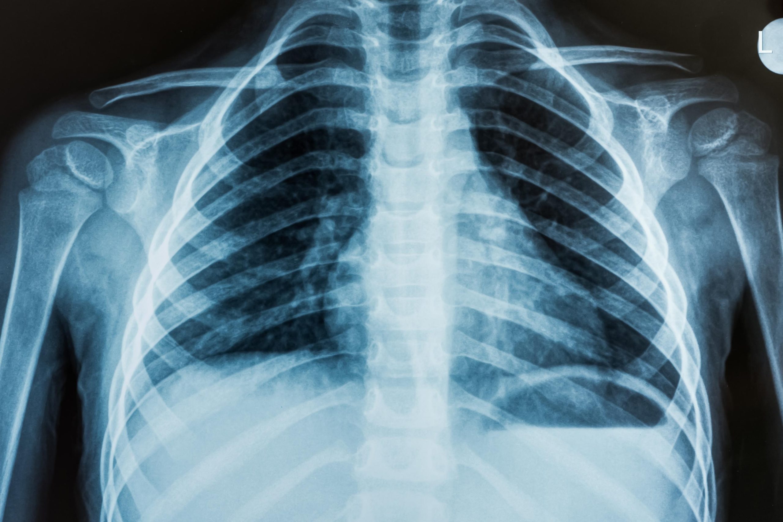
Radiography – captures a single image for subsequent evaluation. Mammography is a specialized form of radiography designed to visualize the internal structures of the breasts.
Fluoroscopy – provides a continuous X-ray image on a monitor, facilitating real-time observation of a procedure or the movement of a contrast agent (“dye”) through the body. Fluoroscopy can expose patients to relatively high radiation doses, particularly during intricate interventional procedures (like the placement of stents or other devices), which necessitate prolonged fluoroscopic monitoring.
CT – involves capturing multiple X-ray images as the detector rotates around the patient’s body. A computer then reconstructs these individual images into cross-sectional views or “slices” of internal organs and tissues. A CT scan typically involves a higher radiation dose compared to standard radiography, as the CT image is derived from numerous individual X-ray projections.
Benefits – Risks
Benefits
The advent of X-rays and the development of CT technology have marked significant milestones in the field of medicine. X-ray imaging is widely acknowledged as an essential diagnostic tool utilized in various examinations and procedures. Its applications include:
– noninvasive and painless assistance in diagnosing diseases and monitoring treatment progress;
– facilitation of medical and surgical planning; and
– guidance for healthcare professionals during the insertion of catheters, stents, or other devices within the body, as well as in the treatment of tumors or the removal of blood clots and other obstructions.
Risks
As with many medical practices, the use of X-ray imaging carries certain risks due to its reliance on ionizing radiation to create body images. Ionizing radiation possesses sufficient energy to potentially harm DNA. The risks associated with exposure to ionizing radiation include:
– a slight increase in the likelihood that an individual exposed to X-rays may develop cancer later in life. (General information regarding cancer detection and treatment can be found through the National Cancer Institute.)
– tissue effects such as cataracts, skin reddening, and hair loss, which typically occur at higher levels of radiation exposure and are uncommon for many imaging procedures. For instance, standard use of a CT scanner or conventional radiography is unlikely to cause tissue effects; however, certain lengthy and complex interventional fluoroscopy procedures may expose the skin to doses that could lead to such effects in specific situations.
Additionally, there is a risk of adverse reactions related to an intravenously administered contrast agent, or “dye,” which is occasionally utilized to enhance image clarity.
The likelihood of developing cancer from radiation exposure during medical imaging is generally minimal and is influenced by:
– radiation dose – The lifetime risk of cancer escalates with higher doses and an increased number of X-ray examinations a patient undergoes.
– patient’s age – The lifetime risk of cancer is greater for individuals who receive X-rays at a younger age compared to those who are older.