The Evolution of Radiology
Radiology is a cornerstone of modern medicine, providing essential insights into the human body’s structure and function. Over the last century, radiology has evolved dramatically, transitioning from the discovery of X-rays to the integration of artificial intelligence (AI) and other cutting-edge technologies. This transformation has not only improved diagnostic accuracy but also revolutionized patient care and treatment outcomes. In this article, we explore the history of radiology, its technological advancements, and the exciting possibilities on the horizon.
The Growth of Radiology: Expanding Imaging Modalities
As the 20th century progressed, radiology expanded beyond X-rays to include new imaging technologies that enhanced diagnostic capabilities.
Fluoroscopy and Contrast Imaging (1920s)
Fluoroscopy, introduced in the early 20th century, allowed doctors to observe real-time images of the internal structures of the body. This technique, which involves the use of a continuous X-ray beam, was invaluable for guiding diagnostic procedures and surgeries. The addition of contrast agents, such as barium for gastrointestinal imaging, further improved the clarity and precision of these procedures.
CT Scanning (1970s)
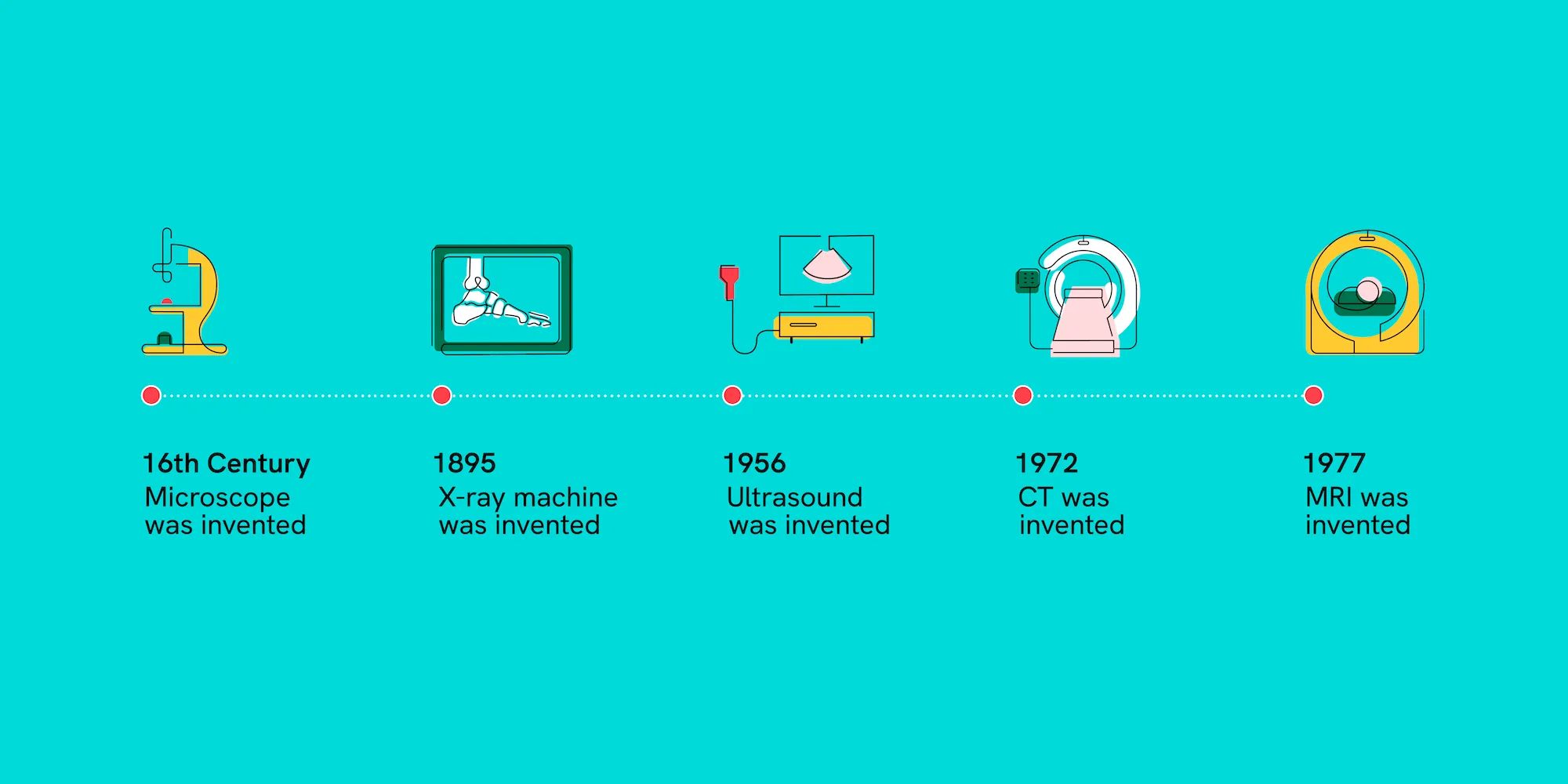
In 1972, Sir Godfrey Hounsfield and Dr. Allan Cormack developed the first computed tomography (CT) scan. This revolutionary imaging technique combined X-ray technology with computer processing to create cross-sectional images of the body, offering more detailed and three-dimensional views than traditional X-rays. CT scans became essential for diagnosing a wide range of conditions, from brain injuries to cancer, and it is now one of the most commonly used imaging modalities worldwide.
MRI (Magnetic Resonance Imaging) (1980s)
MRI, introduced in the early 1980s, marked a major leap forward in non-invasive diagnostic imaging. Unlike X-rays and CT scans, MRI uses powerful magnets and radio waves to produce detailed images of soft tissues, such as the brain, spinal cord, and muscles. MRI’s ability to generate high-resolution, detailed images without exposing patients to radiation made it a valuable tool in detecting conditions such as brain tumors, joint injuries, and heart disease.
Advancements in Image Resolution and Interventional Radiology
As technology advanced, the resolution of images produced by radiology techniques improved dramatically. This increased detail helped clinicians make more accurate diagnoses, leading to better patient outcomes.
Digital Radiography (1990s-Present)
The shift from film-based imaging to digital radiography (DR) began in the 1990s. Digital imaging allowed for faster processing of X-ray images, easier storage and retrieval of patient records, and enhanced image quality. This transition not only improved workflow efficiency but also significantly reduced the amount of radiation patients were exposed to compared to traditional film-based methods.
Interventional Radiology (2000s-Present)
Interventional radiology (IR) represents a paradigm shift in how physicians treat conditions. Using image-guided techniques such as CT, MRI, and ultrasound, interventional radiologists can perform minimally invasive procedures to treat various conditions. These procedures include everything from tumor ablations and angioplasties to biopsies and catheter placements. IR has revolutionized the treatment of cardiovascular diseases, cancer, and other conditions, offering patients quicker recovery times and reduced risks compared to traditional surgeries.
Artificial Intelligence in Radiology: A New Frontier
The most recent and perhaps most exciting development in the field of radiology is the integration of artificial intelligence (AI). AI’s potential to enhance diagnostic accuracy, reduce human error, and increase efficiency has generated significant interest in healthcare.
AI-Assisted Image Interpretation
AI algorithms can now assist radiologists in interpreting medical images by automatically detecting abnormalities, such as tumors, fractures, or infections. These algorithms use deep learning, a type of machine learning, to analyze vast amounts of image data and identify patterns that may be difficult for human eyes to catch. By quickly flagging potential issues, AI reduces the risk of missed diagnoses and allows radiologists to focus on more complex aspects of image interpretation.
One of the most promising applications of AI in radiology is its ability to analyze large datasets, including CT and MRI scans, to assist in the early detection of diseases like cancer, cardiovascular conditions, and neurological disorders. For example, AI is being used to detect early-stage lung cancer in CT scans, which can significantly improve patient survival rates by catching the disease before symptoms arise.