Ultrasound in Cancer Diagnosis: Exploring Its Advantages and Limitations
Ultrasound has long been recognized as a key diagnostic tool in the medical field. Thanks to its real-time, non-invasive imaging capabilities, it plays a crucial role in detecting and monitoring various types of cancers. However, like all diagnostic tools, ultrasound comes with its own set of strengths and limitations. This article delves into how ultrasound is used in the diagnosis of cancer, its benefits, and the potential challenges it poses in clinical practice.
How Ultrasound Assists in Cancer Detection
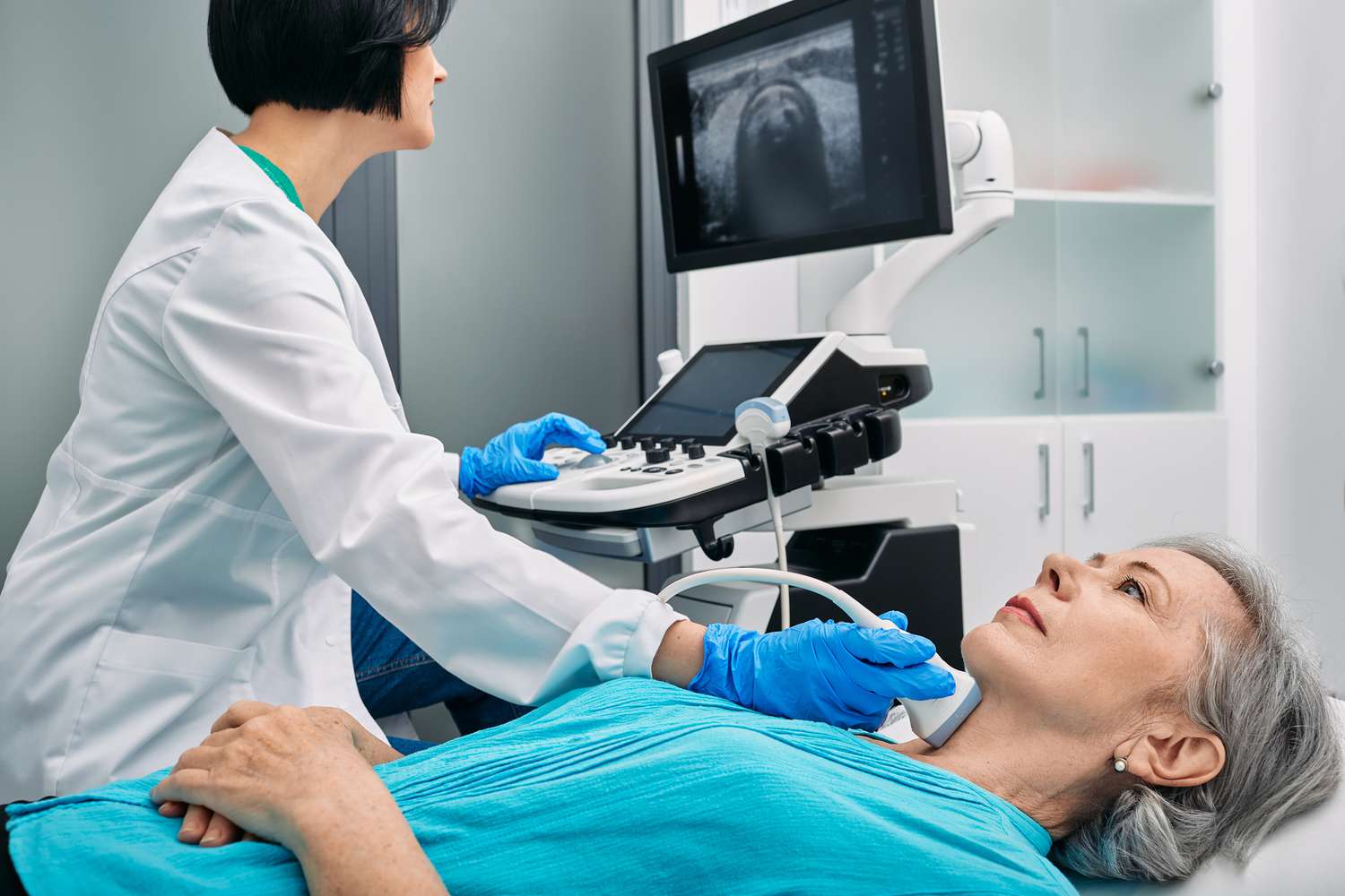
Ultrasound works by emitting high-frequency sound waves through a small probe that is placed on the skin above the area being examined. These sound waves bounce off internal organs and tissues and return to the probe, where they are transformed into visual images. This real-time imaging technique allows medical professionals to detect changes in soft tissues that may indicate the presence of tumors or other abnormalities.
In cancer diagnosis, ultrasound is typically used in conjunction with other imaging modalities, such as CT scans, MRIs, and mammography. It helps healthcare providers assess tumor size, shape, and location, offers guidance for biopsy procedures, and monitors the progress of the disease or response to treatment.
Advantages of Ultrasound in Cancer Diagnosis
- Non-Invasive and Safe
A key benefit of ultrasound is that it is a non-invasive procedure that does not involve radiation, unlike CT scans or X-rays. This makes ultrasound a safer alternative for repeated imaging, especially for vulnerable populations such as pregnant women or young children. It also allows for long-term monitoring of patients without concerns about the cumulative effects of radiation.
- Real-Time Imaging for Precision
Ultrasound provides dynamic, real-time imaging, enabling doctors to observe structures and their movements. This is particularly valuable during procedures like biopsies or needle aspirations, where accurate targeting of the tissue is crucial. The real-time nature of ultrasound helps doctors guide instruments to the tumor site, reducing the risk of harming surrounding tissues and ensuring precise sample collection.
- Affordable and Widely Available
Compared to more expensive imaging methods such as MRI or CT, ultrasound is a cost-effective option. It is less costly to operate and more widely available, with many outpatient clinics and smaller healthcare settings offering ultrasound services. This affordability and accessibility make it a particularly valuable diagnostic tool, especially in low-resource settings.
- Effective for Soft Tissue and Fluid Detection
Ultrasound is highly effective in detecting soft tissue masses and fluid-filled structures, such as cysts and abscesses, which can be indicative of certain cancers. It is commonly used to evaluate tumors in organs such as the liver, kidney, thyroid, and breast, and can also help assess lymph nodes to check for signs of metastasis (cancer spread).
- Guiding Biopsy and Treatment Procedures
Beyond detecting tumors, ultrasound is often used to guide medical procedures like biopsies. It ensures that needles are placed accurately, helping doctors to extract tissue samples from the correct areas. Additionally, ultrasound can assist in procedures such as draining fluid collections or administering treatments like chemotherapy, providing greater precision and minimizing complications.
Challenges and Limitations of Ultrasound in Cancer Diagnosis
- Shallow Penetration of Deep Tissues
A major limitation of ultrasound is its inability to effectively visualize deeper tissues. While it excels at imaging organs close to the surface, such as the breast or thyroid, it struggles to provide clear images of deeper structures like the pancreas or lungs. In cases where tumors are located deep within the body, additional imaging techniques like CT or MRI are often needed for a more comprehensive evaluation.
- Dependence on Operator Expertise
Unlike MRI or CT scans, which produce highly detailed images automatically, ultrasound image quality is highly dependent on the skill and experience of the technician or physician performing the exam. The operator must carefully adjust the probe and interpret the results in real time. This variability can sometimes lead to misinterpretation or missed diagnoses, especially if the ultrasound is not conducted properly.
- Challenges in Differentiating Benign from Malignant Tumors
While ultrasound can effectively detect the presence of a tumor, it often cannot definitively distinguish between benign (non-cancerous) and malignant (cancerous) tumors. Although certain features such as irregular shapes or increased blood flow may suggest malignancy, a biopsy is typically required for a conclusive diagnosis. As such, ultrasound is often used alongside other imaging methods to confirm a diagnosis.
- Lower Sensitivity for Certain Types of Cancer
The sensitivity of ultrasound varies depending on the type of cancer. For example, ultrasound is very effective at detecting breast, liver, and thyroid cancers, but it may not be as useful for identifying cancers of the gastrointestinal tract, lungs, or bones. The visibility of a tumor can be influenced by its size, location, and the composition of surrounding tissues, as well as factors such as the patient’s body fat.
- Difficulty Imaging Certain Anatomical Areas
Ultrasound is not effective at imaging certain body parts, such as bones or air-filled organs like the lungs. These limitations can hinder its ability to detect cancers in these areas. Additionally, ultrasound may struggle to visualize tumors that are encased in dense tissue or are located behind bone or other structures, further reducing its diagnostic utility in such cases.
Ultrasound’s Role in Cancer Screening and Follow-up
Despite these limitations, ultrasound plays an important role in the screening and follow-up of cancer patients. For instance, it is commonly used in breast cancer screening, particularly in women with dense breast tissue, where mammograms may be less effective. Ultrasound can also help monitor patients who are undergoing cancer treatment, allowing doctors to track tumor growth, assess the effectiveness of therapy, and detect any recurrence.
Additionally, ultrasound is useful in evaluating cancer spread to lymph nodes or other nearby structures. In cancers like breast cancer and melanoma, ultrasound can detect enlarged lymph nodes, which may indicate that the cancer has spread (metastasized).