Ultrasound in Obstetrics: Monitoring Life Before Birth
In the realm of modern medicine, few tools have transformed prenatal care as profoundly as the ultrasound machine. From the first flicker of a heartbeat to detailed assessments of fetal anatomy, ultrasound in obstetrics offers a non-invasive window into the womb, allowing healthcare providers to monitor the development of life before birth with astonishing clarity.
A Glimpse into the Womb
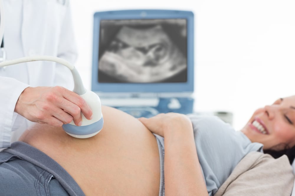
Ultrasound technology uses high-frequency sound waves to create images of internal organs and structures. In obstetrics, this technology enables real-time visualization of the fetus throughout pregnancy. For expectant parents, it is often the first tangible connection to their baby, while for physicians, it is a critical diagnostic tool.
The first ultrasound scan, typically performed around 6 to 9 weeks, confirms pregnancy, estimates gestational age, and can detect multiple fetuses. As the pregnancy progresses, ultrasounds become more detailed, tracking fetal growth, checking for congenital anomalies, and assessing the health of the placenta, amniotic fluid, and uterus.
Key Milestones in Fetal Ultrasound
- First Trimester (0–13 weeks)
- Confirming pregnancy and location (intrauterine vs. ectopic)
- Determining gestational age
- Detecting heartbeat
- Screening for chromosomal conditions via nuchal translucency measurement
- Second Trimester (14–26 weeks)
- Anatomy scan (often at 18–22 weeks): a comprehensive check for structural anomalies
- Monitoring fetal growth and organ development
- Gender identification (if desired)
- Third Trimester (27 weeks onward)
- Assessing fetal position (e.g., breech)
- Estimating fetal weight
- Checking placental position and maturity
- Doppler studies to evaluate blood flow and fetal well-being
Why It Matters
Ultrasound is more than a bonding experience for parents—it’s a vital tool for early detection and intervention. For example:
- Growth restriction can be identified and managed before complications arise.
- Placenta previa, a condition where the placenta covers the cervix, can be monitored to prevent life-threatening bleeding during delivery.
- Congenital anomalies, such as spina bifida or heart defects, may be detected early enough to plan specialized care or even fetal surgery.
Safety and Accessibility
One of the greatest strengths of ultrasound is its safety profile. Unlike X-rays, ultrasound uses sound waves, not ionizing radiation, making it safe for both the mother and the developing fetus. Its portability and cost-effectiveness have also made it an indispensable tool, particularly in remote and low-resource settings.
However, accessibility can still be an issue. In many parts of the world, expectant mothers do not have consistent access to quality prenatal imaging. Global health initiatives continue to push for wider availability of ultrasound technology in underserved areas.
Looking Ahead
Advancements in 3D and 4D ultrasound are enhancing obstetric imaging, offering more detailed and dynamic views of the fetus. These technologies not only provide clearer images but also help diagnose subtle conditions that may go unnoticed with traditional 2D scans.
Artificial intelligence is also beginning to play a role, assisting in image interpretation, reducing operator dependency, and increasing diagnostic accuracy.