Decoding Your Ultrasound Report: What Every Patient Should Know
Undergoing an ultrasound can feel like a big step in your healthcare journey. Whether it’s part of a routine checkup, pregnancy monitoring, or investigating a health concern, this non-invasive imaging technique offers a peek inside your body. But once the test is over, the next challenge is understanding the ultrasound report—often filled with unfamiliar medical terms that can feel confusing or intimidating. Don’t worry! This guide will help you unlock the meaning behind the words and feel more confident about your health.
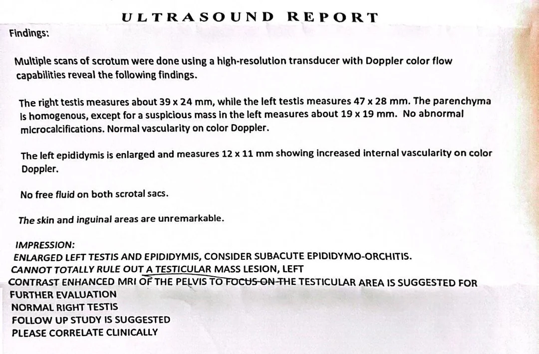
What Exactly Is an Ultrasound Report?
After your ultrasound exam, a specialist called a radiologist examines the images and prepares a report. This document summarizes what the ultrasound revealed about your organs, tissues, or fetus. It highlights any notable findings and provides your doctor with important information needed to guide your care.
Breaking Down Common Ultrasound Terms
Medical jargon can make reports hard to grasp. Here are some terms you might see and what they really mean:
- Echogenicity: This describes how bright or dark something appears on the scan. For instance, a “hyperechoic” area looks brighter and may suggest denser tissue, while a “hypoechoic” spot is darker and could indicate fluid or softer tissue.
- Mass or Nodule: Think of these as lumps or bumps detected during the scan. Reports usually describe their size, shape, and texture to help determine if they’re harmless or if more tests are needed.
- Cyst: A fluid-filled sac that shows up as a dark area on the image. Many cysts are benign and common, but your doctor might want to monitor them.
- Calcifications: These are tiny calcium deposits that appear as bright spots. Depending on their location and pattern, they can be completely normal or sometimes a cause for further investigation.
- Doppler Imaging: This special part of the ultrasound looks at blood flow. It helps your doctor see if your vessels are healthy or if there are any blockages or abnormalities.
How to Make Sense of Your Report
- Focus on the Summary: Look for the section labeled “Impression” or “Conclusion.” This is the radiologist’s main takeaway, usually written in plain language.
- Check for Recommendations: The report may suggest follow-up tests or additional imaging—these are clues about what happens next.
- Talk to Your Doctor: Remember, the ultrasound report is just one part of your overall health picture. Your doctor will explain what it means for you personally.
What Happens If Something Shows Up?
Finding something unusual on an ultrasound can feel worrying, but it’s important to keep perspective. Many findings, like simple cysts or small nodules, are often harmless and just need periodic check-ins. If something requires closer attention, your doctor will guide you through the next steps, which might include more detailed scans or biopsies. Don’t hesitate to ask questions until you feel clear and comfortable.
Tips for Navigating Your Ultrasound Results
- Ask for Your Report and Images: Having your own copy helps you stay informed and get second opinions if you want.
- Prepare Your Questions Ahead of Time: Writing down concerns before your doctor visit can help you cover everything.
- Stay Calm: Many findings are not urgent and monitoring is often the best course.
- Seek Support: It’s perfectly okay to get a second opinion or speak with a specialist for reassurance.