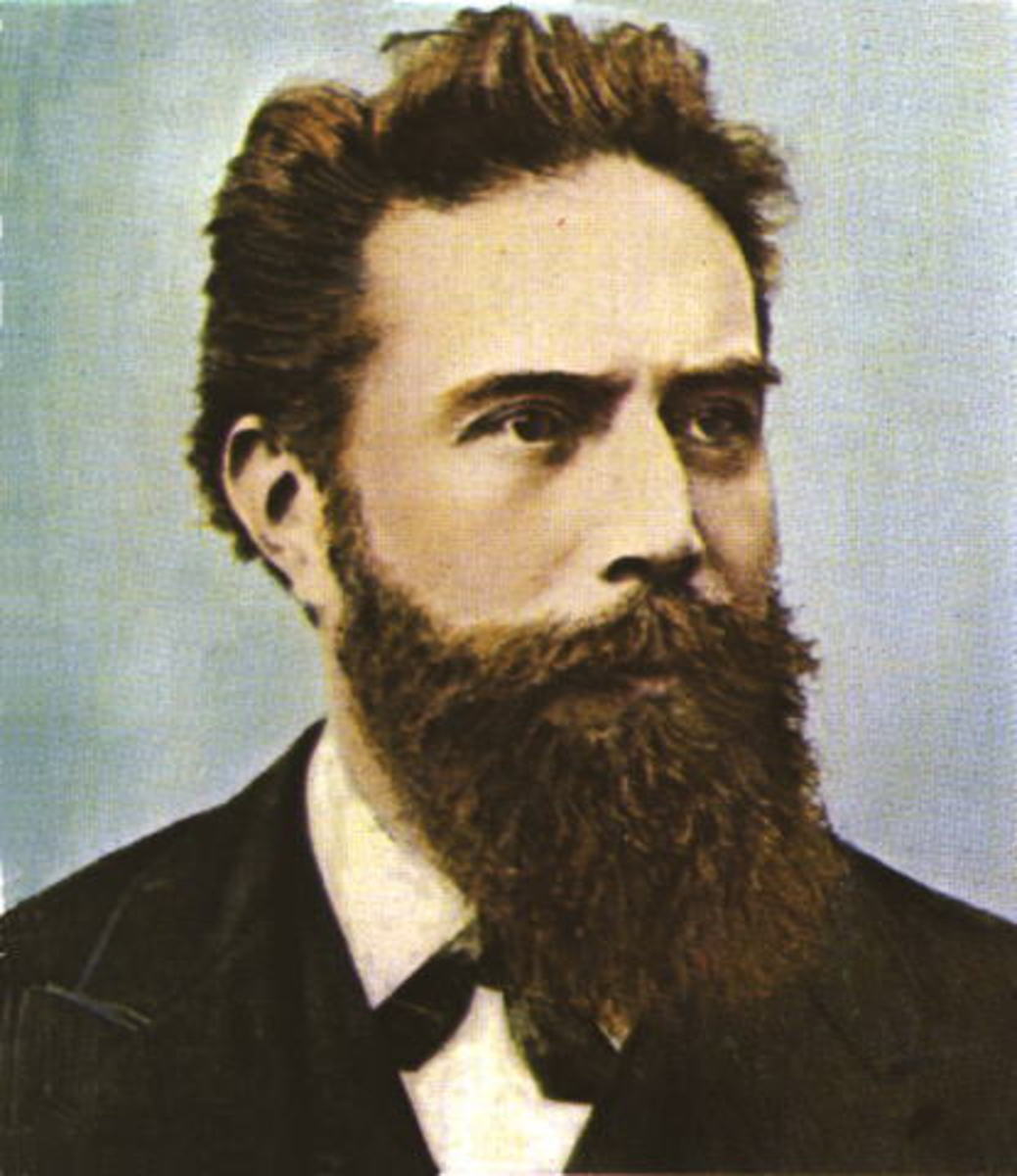
Wilhelm Conrad Röntgen: The Pioneer of Radiology
Radiology traces its beginnings to Wilhelm Conrad Röntgen’s 1895 discovery of X-rays. During his tenure as a physicist, Röntgen focused on a range of scientific inquiries, including piezoelectricity, gas absorption and specific heats, and fluid capillary action. While working with a cathode-ray tube and glass apparatus at the University of Würzburg, he stumbled upon a previously unknown form of rays. These rays could penetrate various materials and cast images on photographic plates. Because the nature of these rays was unknown, he referred to them as “X-rays.”
He shared his breakthrough in a report titled “On a New Kind of Rays,” released on December 28, 1895. News of this phenomenon spread swiftly, particularly among medical professionals. By February 1896, doctors began using X-rays in clinical settings. Röntgen’s pioneering work, which laid the groundwork for modern radiology, earned him the first Nobel Prize in Physics in 1901.
Röntgen’s 1895 introduction of X-rays marked the inception of radiology and shaped its modern evolution.
Evolution of Radiologic Technology
Initial radiographic techniques involved capturing images on glass plates. A major advancement occurred in 1918 when George Eastman, founder of Eastman Kodak, introduced the use of film. Today, radiographic imaging relies on digital technologies and systems like PACS/MIMPS for storing and managing electronic images.
Decades of technological innovation have propelled the field forward. The era following World War II and into the 1950s spurred the creation of ultrasound for medical use. The 1970s saw the introduction of vital imaging modalities still in use today — such as the CAT scan and MRI.
Radiology Milestones Through Time
- 1895
Wilhelm Conrad Röntgen discovers X-rays in Germany. The first X-ray image features his wife’s hand, revealing her skeletal structure and a ring. - 1896
X-ray usage appears in medical practice by January. Around the same time, Antoine-Henri Becquerel in France uncovers radioactivity. - 1914–1918
Radiological tools are employed in World War I field hospitals. - 1918
George Eastman introduces film, replacing glass photographic plates for radiographs. - 1946
Edward Purcell and Felix Bloch, both American physicists, independently discover nuclear magnetic resonance (NMR). - 1955
Ian Donald from Scotland explores ultrasound in gynecology. He collaborates with engineer Tom Brown to build a mobile ultrasound device. - 1961
James Robertson constructs the first single-plane PET scan in the U.S. - 1972
Godfrey Hounsfield, a British engineer, creates the first clinical CT scanner prototype. - 1973
Paul Lauterbur, an American chemist, publishes the first NMR image. - 1975–1980
The introduction of real-time ultrasound machines transforms imaging. - 1977
Raymond Damadian, an American physician, performs the first MRI. - Early 1980s
Hospitals begin installing MRI machines. - 1990s
Ultrasound becomes standard in monitoring fetal development during pregnancy. - 1991
The first functional MRI (fMRI) of the brain is performed by Belliveau and colleagues. - 2000
TIME Magazine names the PET-CT scanner — developed by David Townsend and Ronald Nutt — as the year’s top medical invention. - 2012
November 8 is officially recognized annually as the International Day of Radiology (IDoR). - 2014
The University of Canterbury receives $12 million to develop the first human color X-ray scanner.
The Transformation of Radiology Over the Years
Radiology has come a long way, evolving from glass plate imaging to sophisticated, high-definition digital systems. These innovations have significantly enhanced medical care. Radiology is now supported by modern tools such as PACS, RIS integrations, teleradiology, and advanced Imaging EMRs — now essential in today’s healthcare and medical administration environments.