The Most Vital Equipment in Radiology: The Core of Medical Imaging
Radiology is a key component of contemporary healthcare, granting physicians the ability to examine the body’s internal structures without surgical intervention. Whether identifying bone breaks, detecting tumors, or evaluating internal issues, the practice relies on sophisticated imaging technology. Among all the machines used in this domain, the X-ray machine stands as the most foundational tool in diagnostic radiology.
Why the X-ray Machine Matters Most
Though radiology employs a variety of complex devices—like MRI scanners, CT machines, and ultrasound units—the X-ray machine remains the most commonly utilized and essential piece of equipment in the field. Here’s what makes it so critical:
- Primary Diagnostic Tool: X-rays often serve as the first imaging method used to investigate a wide array of health concerns, including skeletal injuries and respiratory problems. Their speed and availability make them a frontline choice for healthcare providers.
- Affordable and Quick: Compared to more intricate imaging methods like MRI or CT scans, X-rays are both less costly and faster, making them ideal for urgent care and routine clinical use.
- Wide Medical Application: From orthopedic clinics and lung diagnostics to dental care and cancer screening, X-ray machines are applied across numerous branches of medicine.
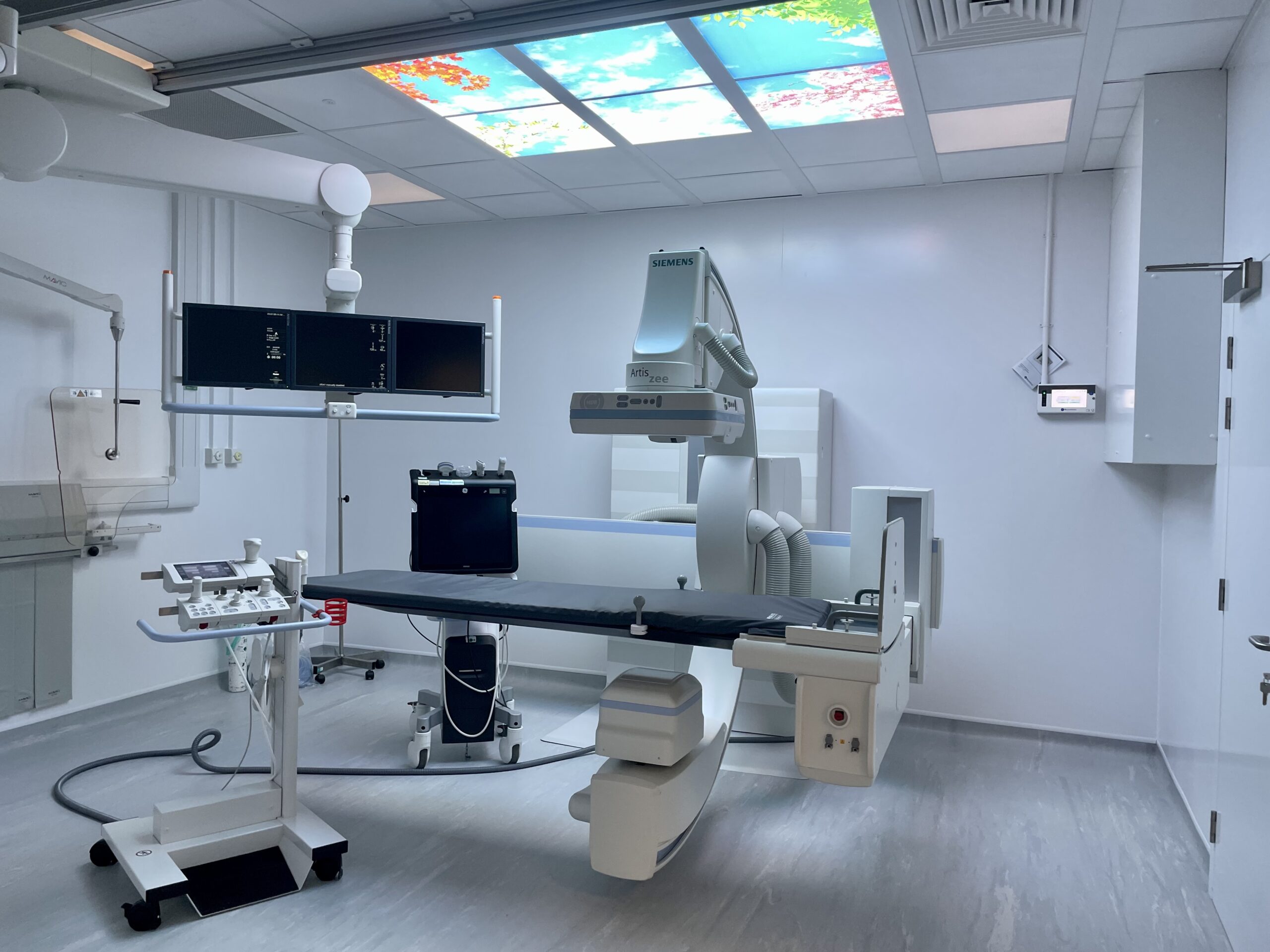
- Essential in Emergency Medicine: In critical care or trauma scenarios, mobile X-ray systems can be quickly deployed to a patient’s bedside for immediate imaging.
How X-ray Imaging Works
X-ray machines function by emitting controlled amounts of radiation that pass through the body to generate internal images. Dense materials, like bone, absorb more radiation and appear white on the resulting image, while softer tissues show up in varying gray tones. These visuals allow doctors to spot issues such as bone fractures, infections, abnormal growths, or fluid accumulation.
Other Key Imaging Devices in Radiology
While X-rays are the cornerstone of most imaging departments, various other machines are indispensable in specific situations:
- CT Scanner (Computed Tomography): Produces in-depth, cross-sectional images of the body, essential for diagnosing conditions such as internal trauma, malignancies, and cerebrovascular accidents.
- MRI (Magnetic Resonance Imaging): Generates detailed views of internal organs and soft tissue using magnetic fields and radio waves—without relying on radiation.
- Ultrasound System: Employs sound waves to produce real-time images, frequently used in pregnancy monitoring, cardiac assessments, and abdominal evaluations.
- Mammogram Unit: A specialized X-ray machine designed for early breast cancer detection.
All of these devices serve important roles, but day-to-day operations in radiology departments typically center around the reliability and usefulness of the X-ray machine.
The Pillar of Diagnostic Imaging
X-ray machines have remained a critical diagnostic asset since their development in the late 1800s. Despite advances in imaging technology, they continue to be a vital part of healthcare in hospitals, outpatient clinics, emergency departments, and even in rural or mobile medical settings.
Ultimately, while radiology is equipped with a wide range of powerful imaging tools, the X-ray machine continues to be the backbone of diagnostic imaging—a vital tool that aids in accurate diagnoses, guides medical interventions, and plays a central role in saving lives.